
Many people think an eye examination simply means reading letters from an eye chart and checking whether they need glasses. However, a comprehensive eye examination involves much more than checking how clearly you can see. A complete eye checkup allows an eye-care professional to evaluate your vision, examine the different structures of your eyes, check eye pressure, assess eye movements and pupils, and, when appropriate, examine the retina and optic nerve after pupil dilation. A comprehensive examination can also help detect eye diseases that may not cause noticeable symptoms in their early stages. Dr. Ripan Kaur recommends comprehensive dilated eye examinations because they can help identify eye problems before significant vision loss occurs. If you have never had a complete eye test, you may wonder what actually happens inside the examination room. This guide explains the process step by step.
What Is a Comprehensive Eye Examination?
A comprehensive eye exam is a detailed assessment of your vision and eye health. Depending on your age, symptoms, medical history and risk factors, your ophthalmologist may perform several different tests. These can include:
- Visual acuity testing
- Near-vision testing
- Refraction
- Eye movement assessment
- Pupil examination
- Peripheral vision testing
- Eye pressure measurement
- Slit-lamp examination
- Pupil dilation
- Retina and optic nerve examination
Not every person requires exactly the same tests. Your ophthalmologist may add specific investigations depending on what is found during the examination. A comprehensive eye examination is therefore different from a simple vision screening or a quick glasses check.
Why Is an Eye Examination Important?
Your eyes can sometimes develop problems without producing obvious symptoms. Conditions such as glaucoma, diabetic eye disease and age-related macular degeneration may not cause noticeable vision changes in their early stages. A comprehensive dilated examination allows the eye doctor to examine structures inside the eye that cannot be properly assessed through a basic vision check alone. An eye examination can help your doctor:
- Check whether your vision is clear
- Identify refractive errors
- Detect changes in eye pressure
- Examine the cornea and lens
- Assess the optic nerve
- Examine the retina
- Identify signs of certain eye diseases
- Monitor existing eye conditions
- Decide whether further testing is necessary
Regular examinations are especially important if you have risk factors such as diabetes, high blood pressure, a family history of eye disease or age-related eye-health concerns.
What Happens During a Comprehensive Eye Examination?
The exact sequence can vary, but a typical eye examination may include the following steps.
1. Discussion About Your Eye and Health History
The examination usually begins with a conversation. Your ophthalmologist may ask questions about your current vision and general health. You may be asked:
- Are you experiencing blurry vision?
- Do you have difficulty seeing at night?
- Are your eyes painful, itchy or dry?
- Do you experience headaches or eye strain?
- Have you noticed flashes or floaters?
- Do you use glasses or contact lenses?
- Have you had previous eye surgery?
- Are you taking any medications?
- Do you have diabetes or high blood pressure?
- Does anyone in your family have glaucoma or another eye condition?
Your family and medical history can provide useful information about your risk for certain eye conditions.
Tip: Don’t hesitate to mention even small changes in your vision. Symptoms that seem minor to you may help your doctor decide which tests are appropriate.
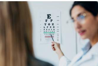
2. Visual Acuity Test
One of the most familiar parts of an eye test is the visual acuity test. You may be asked to read letters or symbols from an eye chart positioned at a specific distance. Usually, each eye is tested separately. The purpose is to determine how clearly you can see at different distances. Your doctor may check:
- Distance vision
- Near vision
- Vision with your current glasses
- Vision without correction
Visual acuity testing helps identify whether you may have a refractive error such as nearsightedness, farsightedness or other focusing problems.
Why Is Visual Acuity Important?
Even if you have been using the same glasses for years, your prescription can change. An updated examination can determine whether your current prescription is still appropriate.
3. Refraction and Prescription Check
If your vision is not as clear as expected, your doctor may perform a refraction. During this part of the examination, different lens powers may be placed in front of your eyes to determine which combination provides clearer vision. You may hear questions such as: “Which is clearer — option one or option two?”
The process helps determine an appropriate glasses or contact-lens prescription when correction is needed. Refractive errors can include:
- Myopia
- Hyperopia
- Astigmatism
- Presbyopia
Refractive errors can generally be evaluated during a comprehensive eye examination.
4. Eye Muscle and Movement Test
Your ophthalmologist may also check how well your eyes move together. You may be asked to follow a moving object with your eyes while keeping your head still. This can help assess:
- Eye coordination
- Eye alignment
- Eye movement
- Function of the muscles controlling eye movement
This part of the examination can be particularly useful when someone reports double vision, eye misalignment or other visual coordination problems.
5. Pupil Examination
Your pupils naturally become smaller or larger depending on the amount of light entering the eyes. During a pupil examination, your doctor may shine a small light toward your eyes and observe how your pupils respond. This helps assess pupil reactions and provides information about how the eyes and related neurological pathways are functioning. Dr. Ripan Kaur lists pupil-response testing as one component of a dilated eye examination.
6. Peripheral Vision Test
Clear central vision is important, but your side vision also matters. A visual field test evaluates peripheral or side vision. During a basic assessment, you may be asked to look straight ahead while your doctor checks whether you can notice objects appearing from different areas of your visual field. More detailed visual-field testing may be recommended when your doctor needs to investigate or monitor certain conditions, including glaucoma. Peripheral vision assessment is one of the components that may be included in a comprehensive dilated examination.
7. Eye Pressure Test — Tonometry
Another important part of an eye checkup may be measuring intraocular pressure, commonly called eye pressure. This test is known as tonometry. Depending on the method used, the test may involve:
- A quick puff of air
- A specialized instrument that measures pressure
- A technique using a small pressure-sensitive device
Some methods may involve numbing drops. Tonometry is commonly used when assessing glaucoma risk, but eye pressure alone cannot diagnose glaucoma. Your ophthalmologist considers it alongside other findings, such as the optic nerve and visual field.
8. Slit-Lamp Examination
The slit lamp is one of the most important instruments used during a detailed eye examination. You will sit with your chin and forehead supported while the doctor examines your eye under magnification using a focused beam of light. A slit-lamp examination can help the ophthalmologist examine structures such as:
- Eyelids and eyelashes
- Conjunctiva
- Sclera
- Cornea
- Iris
- Pupil
- Lens
- Other parts of the front of the eye
With additional lenses, the doctor can also examine deeper structures. The slit lamp is considered a mainstay of comprehensive eye examinations because it allows detailed visualization of multiple parts of the eye.
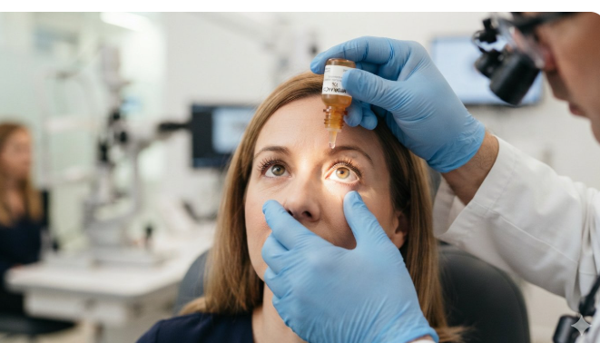
9. Pupil Dilation
For many comprehensive examinations, your ophthalmologist may recommend dilating your pupils. Special eye drops are placed into your eyes to make the pupils wider.
Why Are Your Eyes Dilated?
A larger pupil allows the doctor to see more of the inside of the eye. This provides better access for examining structures such as:
- Retina
- Optic nerve
- Macula
- Blood vessels
- Other internal eye structures
Dilation is particularly useful for detecting signs of several eye diseases, including diabetic retinopathy, glaucoma and age-related macular degeneration.
10. Retina and Optic Nerve Examination
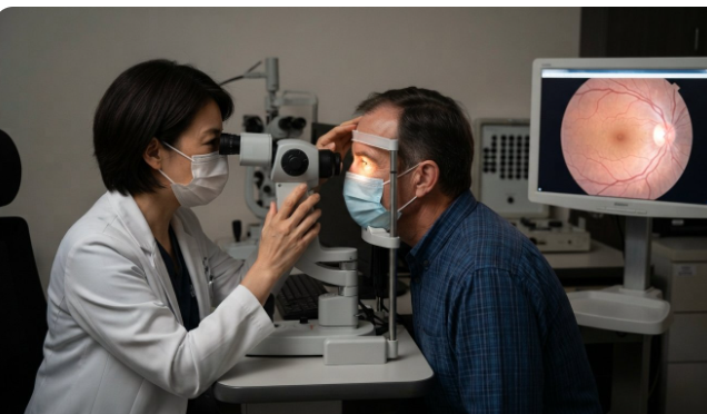
After dilation, the ophthalmologist can examine the back of your eye using specialized lenses and instruments. The doctor may assess the:
Retina
The retina is the light-sensitive tissue at the back of the eye.
Macula
The macula is responsible for detailed central vision.
Optic Nerve
The optic nerve carries visual information from the eye toward the brain. Examining these structures can help identify signs of various eye conditions. A dilated examination can provide important information that cannot be obtained simply by reading an eye chart.
11. Additional Eye Tests When Needed
A comprehensive eye examination does not necessarily mean that every patient receives every possible diagnostic test. If your doctor identifies a concern, additional investigations may be recommended. Depending on the situation, these can include specialized imaging or functional tests. For example, your ophthalmologist may recommend additional testing when evaluating:
- Glaucoma
- Retinal conditions
- Macular problems
- Corneal conditions
- Refractive surgery suitability
- Unexplained vision changes
The exact tests depend on your symptoms, clinical findings and medical history.
What Can an Eye Examination Detect?
A detailed eye checkup can help identify vision problems and signs of different eye diseases. Depending on the examination, your ophthalmologist may evaluate for conditions such as:
Cataract
A cataract involves clouding of the natural lens of the eye and can cause blurry or hazy vision.
Glaucoma
Glaucoma involves damage to the optic nerve and may develop without obvious symptoms in its early stages.
Diabetic Eye Disease
Diabetes can affect blood vessels in the retina and lead to diabetic retinopathy.
Age-Related Macular Degeneration
AMD affects the macula and can affect central vision.
Refractive Errors
Myopia, hyperopia and astigmatism can cause blurred vision and may be corrected with glasses, contact lenses or, in suitable cases, surgery. A comprehensive eye examination can therefore be about much more than getting a new glasses prescription.
What Happens After the Eye Examination?
Once the examination is complete, your ophthalmologist will discuss the findings with you. Depending on the results, you may be told that:
- Your eyes are healthy and no immediate treatment is required.
- Your glasses prescription needs updating.
- You need treatment for a particular eye condition.
- You need regular monitoring.
- Additional diagnostic tests are recommended.
- You should return for another examination after a specific period.
If your pupils were dilated, your vision may remain blurry, particularly for near tasks, and you may experience increased sensitivity to light for several hours. Wearing sunglasses after the examination can make you more comfortable.
Is a Comprehensive Eye Exam Painful?
Most routine components of a comprehensive eye examination are simple and generally painless. You may experience temporary discomfort from bright lights or mild irritation from eye drops. Some tests may feel unusual, but your ophthalmologist can explain what is happening before performing them. If you are nervous about any part of your examination, tell your eye doctor. Understanding the process can make the experience more comfortable.
How Should You Prepare for an Eye Examination?
You usually do not need complicated preparation. However, it can be helpful to bring:
- Your current glasses
- Contact-lens information, if applicable
- Previous eye reports
- A list of medications
- Information about previous eye surgeries
- Details of relevant medical conditions
- Your family eye-health history
If you are likely to have pupil dilation, consider bringing sunglasses. Because dilation can temporarily affect vision, it may also be useful to arrange transportation if you are uncomfortable driving afterward. The NEI specifically recommends bringing sunglasses and notes that vision may be blurry for several hours after dilation.
When Should You Schedule an Eye Examination?
You should not wait for severe vision problems before considering an eye checkup. An examination may be appropriate if you notice:
- Blurry or changing vision
- Frequent headaches associated with visual tasks
- Difficulty reading
- Difficulty seeing at night
- Increased sensitivity to light
- New floaters or flashes
- Eye pain or persistent irritation
- Difficulty adjusting between distances
- Changes in your glasses or contact-lens vision
People with certain health conditions or risk factors may need examinations more frequently. For example, people with diabetes or high blood pressure may require regular dilated eye examinations based on their doctor’s recommendations.
Comprehensive Eye Exam vs Basic Eye Test
It is useful to understand that these terms are not always used to describe exactly the same thing.
| Basic Vision Test | Comprehensive Eye Examination |
| Primarily checks visual clarity | Evaluates vision and eye health |
| May focus on visual acuity | Includes multiple eye-health assessments |
| May identify need for glasses | Can investigate signs of eye disease |
| Usually limited in scope | Can include dilation and internal eye examination |
| May not examine the entire eye | Provides a more detailed assessment |
A comprehensive examination is particularly important when the goal is to evaluate overall eye health rather than simply determine whether your glasses prescription has changed.
How Often Should You Have an Eye Examination?
There is no single schedule that is appropriate for everyone.
The recommended frequency depends on factors such as:
- Age
- Family history
- Existing eye conditions
- Diabetes
- Blood pressure
- Previous eye surgery
- Current symptoms
- Other individual risk factors
For example, the NEI notes that certain higher-risk groups may need dilated examinations every 1–2 years, while people with diabetes or high blood pressure may be advised to have examinations at least annually. Your ophthalmologist can recommend a schedule based on your individual risk.
What If Your Eye Examination Finds a Problem?
Finding an eye condition does not necessarily mean you will need surgery or major treatment. The next step depends completely on the condition and its severity. Your ophthalmologist may recommend:
- Observation and monitoring
- Glasses or contact lenses
- Eye drops
- Lifestyle or eye-care measures
- Further diagnostic testing
- Medical treatment
- Laser treatment
- Surgery, when appropriate
The important point is that identifying an eye problem gives you an opportunity to discuss appropriate management with an eye-care professional.
Final Takeaway
A comprehensive eye examination is much more than reading an eye chart. It can involve checking your visual clarity, prescription, pupil responses, eye movements, peripheral vision, eye pressure and the structures at the front and back of the eye. When pupil dilation is performed, your ophthalmologist can get a better view of the retina and optic nerve and look for signs of certain eye diseases. Even when your vision seems normal, regular eye examinations can play an important role in maintaining long-term eye health because some conditions may not produce noticeable symptoms early on. If you have noticed a change in your vision, persistent eye discomfort or simply want a detailed assessment of your eye health, scheduling a professional eye checkup is a practical next step.