
Before any eye surgery, your ophthalmologist needs to understand the condition of your eyes in detail. This is why tests before eye surgery are an important part of the treatment process. These tests are not simply about checking whether you can see clearly. Depending on the planned procedure, your doctor may evaluate your vision, cornea, eye pressure, lens, retina, optic nerve, tear film and the overall health of your eyes. For cataract surgery, measurements may be needed to calculate the appropriate intraocular lens (IOL). For LASIK, detailed corneal measurements help assess whether the cornea is suitable for laser vision correction. Other surgeries may require additional imaging or specialized tests. It is also important to understand that not every patient needs every possible test. The tests selected depend on your eye condition, planned surgery, medical history and findings during the examination. The American Academy of Ophthalmology notes that routine general medical testing before eye surgery is not automatically necessary for every patient and should be guided by individual history and examination. In this guide, we explain the most common preoperative eye tests, why they are performed and how they can help your ophthalmologist plan treatment.
Why Are Eye Tests Done Before Surgery?
The main purpose of an eye examination before surgery is to collect enough information to plan the procedure safely and appropriately. Preoperative testing may help your doctor:
- Confirm the diagnosis
- Measure your current vision
- Identify other eye conditions
- Assess the health of the cornea
- Measure eye pressure
- Examine the lens
- Check the retina and optic nerve
- Measure the eye for an IOL
- Evaluate the tear film and ocular surface
- Identify conditions that may affect surgical suitability
- Plan the surgical technique
- Discuss realistic visual expectations
The exact testing process varies according to the type of surgery.
1. Medical and Eye History
Before performing diagnostic tests, your ophthalmologist will usually ask questions about your medical and eye history. This may include:
Previous Eye Conditions
Your doctor may ask whether you have had:
- Cataracts
- Glaucoma
- Dry eye
- Corneal disease
- Retinal problems
- Eye infections
- Eye injuries
- Previous eye surgery
Previous Eye Procedures
Tell your ophthalmologist if you have previously undergone:
- LASIK
- PRK
- Other laser eye procedures
- Cataract surgery
- Corneal surgery
- Retinal surgery
Previous refractive surgery can be particularly important when planning later procedures.
Contact Lens History
If you wear contact lenses, your doctor may ask about the type and duration of use. Contact lenses can temporarily alter corneal shape, which is especially relevant when accurate measurements are required for refractive surgery.
2. Visual Acuity Test
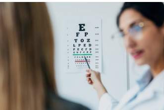
A visual acuity test is one of the most familiar tests performed during an eye examination. You are usually asked to read letters or symbols from an eye chart.
The test may assess:
- Distance vision
- Near vision
- Vision with glasses
- Vision without glasses
For some procedures, your doctor may perform additional refraction testing to determine the exact prescription. Visual acuity is only one part of the evaluation. A person can have relatively good vision while still having an underlying eye condition that needs attention. The National Eye Institute includes visual acuity testing as part of a comprehensive dilated eye examination.
3. Refraction Test
A refraction test determines the optical prescription needed to correct your vision. Your ophthalmologist or optometrist may use different lenses or an automated instrument to determine whether you have:
- Myopia
- Hyperopia
- Astigmatism
- Other refractive errors
For refractive procedures such as LASIK, accurate refraction is an important part of the preoperative assessment. The American Academy of Ophthalmology’s refractive-surgery guidance includes distance and near visual acuity with and without correction and, when appropriate, manifest or cycloplegic refraction.
4. Slit-Lamp Examination
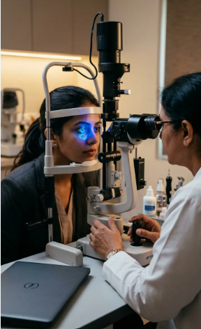
A slit-lamp examination allows the ophthalmologist to examine the structures at the front of the eye under magnification. The doctor can assess structures such as:
- Eyelids
- Conjunctiva
- Cornea
- Anterior chamber
- Iris
- Lens
This examination can help identify conditions that could affect surgical planning. For cataract evaluation, the ophthalmologist may assess the severity of the cataract as well as the health of the cornea, anterior chamber, lens and other structures.
What Happens During a Slit-Lamp Test?
You place your chin and forehead against the supports of the instrument and look toward a target. A narrow beam of light is directed into the eye while the doctor examines the structures through a microscope. It is generally a quick and non-invasive examination.
5. Eye Pressure Test — Tonometry
Tonometry measures the pressure inside the eye, known as intraocular pressure (IOP). It is particularly relevant when evaluating patients for conditions such as glaucoma. During the test, your eye pressure may be measured using an instrument that gently touches the eye or uses an air puff, depending on the equipment.
Why Is Eye Pressure Checked Before Surgery?
Knowing your eye pressure can help your ophthalmologist identify previously undiagnosed or poorly controlled pressure-related problems. Eye pressure is only one component of glaucoma assessment; doctors also consider the optic nerve, visual field and other findings.
6. Dilated Eye Examination
A dilated eye examination allows the ophthalmologist to examine structures at the back of the eye more thoroughly. Dilating drops enlarge the pupil. Your doctor can then examine areas such as:
- Retina
- Macula
- Optic nerve
- Blood vessels
- Posterior eye structures
The National Eye Institute describes visual acuity, visual field, pupil response and tonometry among components that may be included in a comprehensive eye examination, with dilation allowing examination of the back of the eye. For cataract surgery planning, the health of the retina and optic nerve is important because cataract removal cannot correct vision problems caused by unrelated retinal or optic-nerve disease.
7. Pupil Size Measurement
Pupil size may be measured during certain preoperative evaluations. This can be especially relevant in refractive surgery planning because visual symptoms such as glare and halos can be influenced by several factors, including pupil size and optical characteristics of the eye. The FDA’s refractive-surgery testing information includes pupil-size assessment as part of preoperative evaluation. Your doctor may consider the measurement together with your symptoms, eye health and other test results.
8. Corneal Topography
Corneal topography creates a detailed map of the shape and curvature of the cornea. The cornea is the transparent front surface of the eye and plays a major role in focusing light. Topography can help evaluate:
- Corneal curvature
- Astigmatism
- Irregular corneal shape
- Certain corneal abnormalities
It is particularly important before refractive procedures such as LASIK. The American Academy of Ophthalmology’s EyeWiki explains that preoperative topography can help evaluate corneal shape and identify conditions such as keratoconus, irregular astigmatism and contact-lens-induced corneal changes.
9. Corneal Tomography
Corneal tomography goes beyond measuring the front surface of the cornea. It can create a more detailed three-dimensional assessment of the cornea, including information about its anterior and posterior surfaces and thickness distribution. This can be useful when evaluating patients for refractive surgery.
Topography vs Tomography
Topography: Primarily maps the surface shape of the cornea.
Tomography: Provides more comprehensive three-dimensional information about the cornea.
The exact technology used depends on the clinic and the type of surgery being considered.
10. Pachymetry — Measuring Corneal Thickness
Pachymetry measures the thickness of the cornea. This test is particularly important before corneal laser procedures because the surgeon needs to understand the available corneal tissue and overall corneal structure. The American Academy of Ophthalmology includes central corneal thickness measurement in the preoperative evaluation for refractive surgery.
Why Does Corneal Thickness Matter?
LASIK and other corneal procedures alter corneal tissue. Therefore, your surgeon needs accurate information about corneal thickness when assessing suitability and planning treatment. A thin or structurally abnormal cornea may change the recommended treatment approach.
11. Keratometry
Keratometry measures the curvature and refractive power of the cornea. It can provide information about:
- Corneal curvature
- Astigmatism
- Corneal refractive power
Keratometry is particularly important during cataract surgery planning because corneal measurements contribute to the calculation of the appropriate IOL power.
12. Biometry Before Cataract Surgery
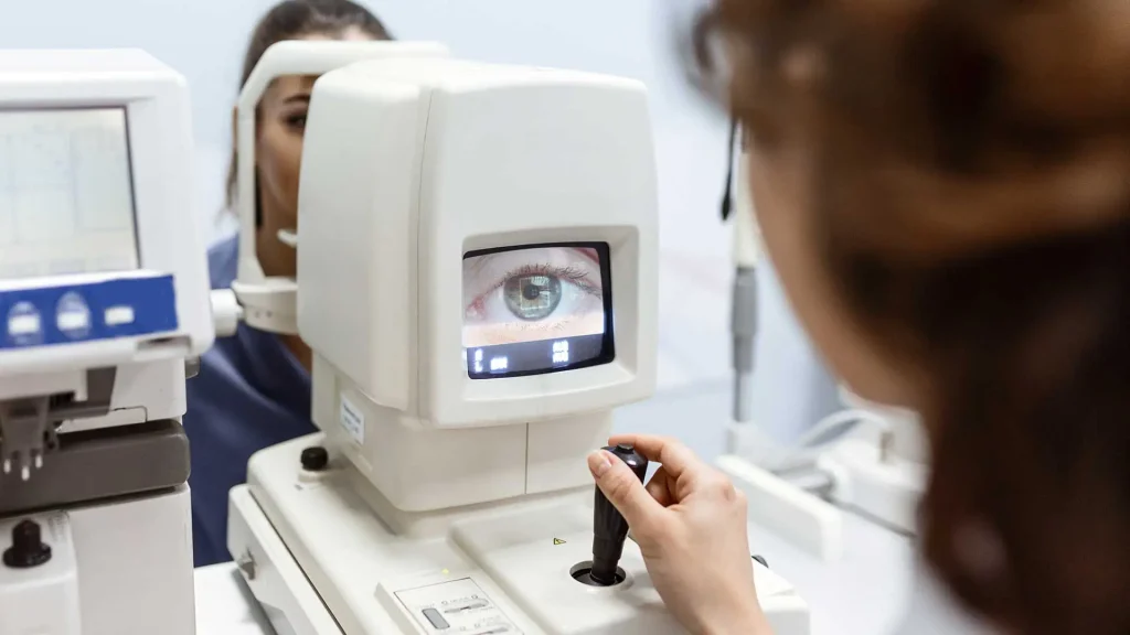
Eye biometry is performed before cataract surgery to measure the eye and help determine the appropriate intraocular lens (IOL) power. If you are having cataract surgery, one of the most important preoperative tests is ocular biometry. Biometry measures different dimensions of the eye that are used to calculate the power of the artificial intraocular lens. The National Eye Institute states that eye measurements are performed before cataract surgery to help choose the appropriate IOL. Measurements may include:
- Axial length
- Corneal curvature
- Anterior chamber depth
- Other eye dimensions depending on the device
EyeWiki notes that axial length, corneal refractive power and anterior chamber depth are among the measurements used for IOL power calculation.
13. IOL Power Calculation
Biometry is closely connected with IOL power calculation. During cataract surgery, the cloudy natural lens is removed and replaced with an artificial intraocular lens. The surgeon needs to select an IOL with an appropriate power based on the measurements of your eye and the intended postoperative refractive target. This is why accurate measurements before cataract surgery are so important. Your ophthalmologist may also discuss different IOL options depending on your visual needs and eye health.
14. Ocular Surface and Dry-Eye Evaluation
The health of the eye surface is important before surgery. Your doctor may look for:
- Dry eye
- Excessive tearing
- Blepharitis
- Eyelid problems
- Corneal surface irregularities
- Tear-film instability
This is particularly relevant before refractive surgery. The American Academy of Ophthalmology’s preoperative LASIK evaluation includes assessment of the tear film and ocular surface. If significant dry eye is identified, your ophthalmologist may recommend treatment before proceeding with certain procedures.
15. Eye Muscle and Alignment Examination
An eye examination may also include evaluation of:
- Eye movement
- Eye alignment
- Coordination between the eyes
The National Eye Institute lists an eye-muscle-function assessment as part of a comprehensive dilated eye examination. This can provide additional information about how the eyes work together.
16. Visual Field Test
A visual field test checks your peripheral or side vision. You focus on a central target and indicate when you see lights or objects appearing in different areas. This test can be particularly relevant when there is concern about glaucoma or optic-nerve function. The National Eye Institute includes visual field testing among components that may be used during comprehensive eye evaluation. Not every patient undergoing every type of eye surgery will require a visual field test.
17. Optical Coherence Tomography — OCT
Optical coherence tomography (OCT) is a non-invasive imaging technique that can create detailed cross-sectional images of eye structures. Depending on the clinical situation, OCT may be used to evaluate:
- Retina
- Macula
- Optic nerve
- Retinal nerve fiber layer
For example, if the doctor suspects a retinal or optic-nerve condition, additional imaging may be useful during preoperative assessment. EyeWiki notes that OCT of the retina and retinal nerve fiber layer may be included as additional imaging in selected cataract cases.
Is OCT Required Before Every Eye Surgery?
No. OCT is generally performed when the ophthalmologist believes it can provide useful information based on the patient’s condition and planned procedure.
18. Retina Examination
A detailed retinal examination may be especially important when the patient has:
- Diabetes
- Previous retinal problems
- Unexplained vision loss
- Macular disease
- High-risk medical history
Before cataract surgery, examining the retina helps the doctor understand whether other retinal conditions could affect the expected visual outcome.
19. Glare or Contrast Testing
Some patients may undergo additional visual-function testing if their symptoms suggest that standard visual acuity does not tell the entire story. For example, cataract evaluation may sometimes include glare or brightness-acuity testing. This can help determine how cataract-related lens opacity affects vision in situations such as bright light or night-driving conditions. These tests are not necessarily performed for every patient.
20. Anterior Segment Imaging
In selected patients, doctors may use specialized imaging to examine structures at the front of the eye. This can provide information about:
- Cornea
- Anterior chamber
- Iris
- Lens
- Other anterior-segment structures
The need for this type of testing depends on the condition being evaluated.
21. Tests for Specific Eye Conditions
Additional testing may be recommended when your doctor suspects a particular condition. For example:
Before Glaucoma Surgery
Your doctor may evaluate:
- Eye pressure
- Optic nerve
- Visual field
- Angle structures
- Retinal nerve fiber layer
Before Corneal Surgery
Testing may include:
- Corneal topography
- Corneal tomography
- Pachymetry
- Slit-lamp examination
Before Retinal Surgery
Additional retinal imaging or ultrasound may be required depending on the condition.
Before LASIK
Preoperative evaluation can include:
- Visual acuity
- Refraction
- Corneal topography/tomography
- Corneal thickness
- Tear-film evaluation
- Eye alignment
- Detailed ocular examination
The AAO’s current refractive-surgery guidance specifically lists these elements as part of the comprehensive preoperative assessment.
Do You Need Blood Tests Before Eye Surgery?
This is one of the most common questions patients ask. Not every eye-surgery patient needs routine blood tests. The American Academy of Ophthalmology notes that routine preoperative medical testing is not necessary for many patients undergoing procedures such as cataract surgery. Additional tests should be based on the patient’s medical history and physical assessment. For example, a doctor may consider additional testing when a patient has conditions such as:
- Diabetes
- Heart disease
- Kidney disease
- Blood-pressure problems
- Other significant medical conditions
The specific requirements depend on the patient’s health and the type of surgery.
Why Your Medical History Can Change the Tests You Need
Two patients having the same eye surgery may not undergo exactly the same testing. For example:
Patient A: Healthy adult with no significant medical history.
Patient B: Patient with diabetes, glaucoma and previous eye surgery.
The second patient may need additional evaluation because their medical and eye history can affect surgical planning.This is why preoperative eye tests are personalized rather than being a single fixed checklist for everyone.
What Happens If a Test Shows a Problem?
An abnormal test result does not automatically mean that surgery is impossible. Your ophthalmologist may:
- Repeat the test.
- Perform another diagnostic examination.
- Treat an underlying eye condition.
- Modify the surgical plan.
- Recommend a different procedure.
- Delay surgery until the condition is stable.
- Discuss whether the expected benefits outweigh the risks.
For example, significant ocular-surface disease may need to be addressed before refractive surgery, while abnormal corneal topography may affect whether laser vision correction is appropriate.
How to Prepare for Your Preoperative Eye Tests
You can make your examination more useful by preparing some basic information.
Bring Your Previous Reports
If you have previous eye-surgery records, prescriptions, scans or reports, take them with you.
Bring Your Current Glasses
Your current glasses and prescription can help your doctor understand your vision history.
Bring Your Medication List
Include prescription medicines, eye drops, supplements and other medications.
Tell Your Doctor About Contact Lenses
Mention what type of lenses you use and how frequently you wear them. For refractive surgery, you may be asked to stop wearing lenses for a specific period before measurements. The required time varies by lens type and procedure.
Mention Previous LASIK or Other Surgery
Previous procedures can affect measurements and surgical planning.
Do All Eye Surgeries Require the Same Tests?
No. This is one of the most important things to remember.
| Surgery | Common Evaluation/Tests |
| Cataract Surgery | Visual acuity, refraction, slit lamp, dilated exam, biometry, corneal measurements |
| LASIK | Visual acuity, refraction, corneal topography/tomography, pachymetry, ocular-surface evaluation |
| Glaucoma Surgery | Eye pressure, optic-nerve assessment, visual field and other glaucoma-specific testing |
| Corneal Surgery | Slit lamp, topography/tomography, pachymetry and other corneal assessments |
| Retinal Surgery | Dilated retinal examination and imaging when indicated |
This table is a general overview. Your ophthalmologist may order fewer, additional or different tests depending on your individual condition.
Final Takeaway
The tests before eye surgery are an important part of individualized surgical planning. Your ophthalmologist may assess your vision, prescription, cornea, eye pressure, lens, retina, optic nerve and ocular surface. Depending on the procedure, specialized tests such as corneal topography, tomography, pachymetry, biometry or OCT may also be recommended. For cataract surgery, accurate eye measurements help calculate the appropriate IOL power. For LASIK and other refractive procedures, corneal measurements and ocular-surface evaluation help determine whether the procedure is appropriate and assist with treatment planning. Remember that there is no single preoperative test list that applies to every patient. The right testing plan depends on your eye condition, medical history and the surgery being considered. If you have been advised to undergo eye surgery, discuss your test results and treatment plan with your ophthalmologist before proceeding.